Development & Reproduction
Korean Society of Developmental Biology
Research article

Styrene Cytotoxicity in Testicular Leydig Cells In Vitro

Jin-Yong Chung1,*https://orcid.org/0000-0001-8149-584X, Ji-Eun Park1,*https://orcid.org/0000-0001-5110-0501, Yoon-Jae Kim1https://orcid.org/0000-0001-5378-9947, Seung-Jin Lee2https://orcid.org/0000-0001-7109-6423, Wook-Joon Yu2https://orcid.org/0000-0003-3844-0274, Jong-Min Kim1,https://orcid.org/0000-0002-6201-1739
1Department of Anatomy and Cell Biology, College of Medicine, Dong-A University, Busan 49201, Korea
2Developmental and Reproductive Toxicology Research Group, Korea Institute of Toxicology, Daejeon 34114, Korea
Corresponding author Jong-Min Kim, Department of Anatomy and Cell Biology, College of Medicine, Dong-A University, Busan 49201, Korea. Tel: +82-51-240-2792, E-mail: jmkim7@dau.ac.kr

These authors contributed equally to this work.

© Copyright 2022 The Korean Society of Developmental Biology. This is an Open-Access article distributed under the terms of the Creative Commons Attribution Non-Commercial License (http://creative-commons.org/licenses/by-nc/3.0/) which permits unrestricted non-commercial use, distribution, and reproduction in any medium, provided the original work is properly cited.

Received: Jun 10, 2022 ; Revised: Aug 03, 2022 ; Accepted: Aug 22, 2022

Published Online: Sep 30, 2022

Abstract

Styrene is the precursor of polystyrene. Human exposure to styrene could occur in occupational and residential settings and via food intake. Styrene is metabolized to styrene-7,8-oxide by cytochrome P450 enzyme. In the present study, we investigated the cytotoxicity mediated by styrene and styrene-7,8-oxide in TM3 testicular Leydig cells in vitro. We first monitored the nuclear fragmentation in Leydig cells after exposure to styrene or styrene-7,8-oxide. Hoechst 33258 cell staining showed that styrene exposure in TM3 Leydig cells did not exhibit nuclear fragmentation at any concentration. In contrast, nuclear fragmentation was seen in styrene-7,8-oxide-exposed cells. These results indicate that cytotoxicity-mediated cell death in Leydig cells is more susceptible to styrene-7,8-oxide than to styrene. Following styrene treatment, procaspase-3 and XIAP protein levels did not show significant changes, and cleaved (active) forms of caspase-3 were not detected. Consistent with the western blot results, the active forms of caspase-3 and XIAP proteins were not prominently altered in the cytoplasm of cells treated with styrene. In contrast to styrene, styrene-7,8-oxide induced cell death in an apoptotic fashion, as seen in caspase-3 activation and increased the expression of XIAP proteins. Taken together, the results obtained in this study demonstrate a fundamental idea that Leydig cells are capable of protecting themselves from cytotoxicity-mediated apoptosis as a result of styrene exposure in vitro. It remains unclear whether the steroid-producing function, i.e., steroidogenesis, of Leydig cells is also unaffected by exposure to styrene. Therefore, further studies are needed to elucidate the endocrine disrupting potential of styrene in Leydig cells.

Keywords: Styrene; Styrene-7,8-oxide; Cytotoxicity; Apoptosis; Caspase-3; Leydig cells

INTRODUCTION

Styrene, an organic compound with the chemical formula C8H8, is the precursor of polystyrene and is primarily produced via the catalytic dehydrogenation of ethylbenzene. Approximately 35 million tons of styrene were produced in 2018, with increasing amounts synthesized annually to manufacture products such as plastic, fiberglass, automobile parts, and food containers. Thus, human exposure to styrene could occur in occupational and residential settings and via food intake. Styrene is metabolized to styrene-7,8-oxide by cytochrome P450 2E1 in the human liver (Guengerich et al., 1991). Both styrene and styrene-7,8-oxide are considered toxic, mutagenic, and carcinogenic. In particular, styrene exhibits neurotoxic properties, and exposure to styrene has been correlated with dysfunctions of sensory organs, such as disorders of vision or hearing (Möller et al., 1990; Gong et al., 2002; Corsi et al., 2007; Gopal et al., 2011; Sliwinska-Kowalska et al., 2020).

Several environmental chemicals exhibit reproductive toxicity in mammalian testes (Akingbemi et al., 2004; Chung et al., 2007; Chung et al., 2011; Traore et al., 2016). Toxicological targets in the testis include germ cells, Sertoli cells, Leydig cells, or all these cells. Among these cell types, Leydig cells, located in the interstitial compartment between seminiferous tubules, produce testosterone, which is critically important for maintaining male homeostasis and reproduction (i.e., spermatogenesis) (Zirkin, 1998). Therefore, potential cellular damage mediated via a specific environmental toxicant could result in Leydig cell death, thereby hindering sperm production (Chung et al., 2011). In addition, given that Leydig cells possess a series of steroidogenic machinery (proteins), including steroidogenic acute regulatory proteins, cytochrome P450 side chain cleavage, and 3β-hydroxysteroid dehydrogenase, the potential dysregulation of these proteins following exposure to environmental chemicals could seriously impact testosterone production and spermatogenesis (Chung et al., 2011; Abdel-Maksoud et al., 2019).
dr-26-3-99-g1
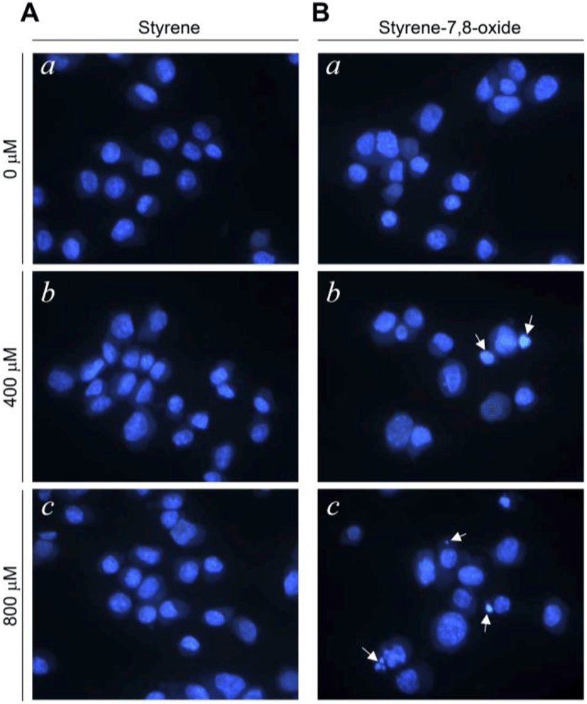
Fig. 1. Effects of styrene or styrene-7,8-oxide on nuclear morphology in TM3 Leydig cells. Cells were treated with 0 (a), 400 (b), and 800 (c) μM styrene (A) or styrene-7,8-oxide (B) for 48 h. Nuclear morphology stained by Hoechst 33258. Arrows point to fragmented nuclei. Original magnification: ×800.
Download Original Figure

Accordingly, in the present study, we investigated the cytotoxicity mediated by styrene and styrene-7,8-oxide in TM3 testicular Leydig cells in vitro. To evaluate the cytotoxicity at cellular and molecular levels, we examined apoptosis morphologically and measured the altered expression of apoptosis-related proteins after exposure to styrene or styrene-7,8-oxide. The purpose of this study was to determine whether styrene or its metabolite (styrene-7,8-oxide) could potentially impact Leydig cell fate.

MATERIALS AND METHODS

1. Reagents and antibodies

Styrene and styrene-7,8-oxide were obtained from Tokyo Chemical Industry (Tokyo, Japan). Hoechst 33258, paraformaldehyde, propidium iodide (PI), and Tween-20 were from Sigma-Aldrich (St. Louis, MO, USA). Anti-caspase-3 was from Santa Cruz Biotech (Santa Cruz, CA, USA). Anti-cleaved caspase-3 was from Cell Signaling Technology (Beverly, MA, USA). Anti-XIAP was from Trevigen (Gaithersburg, MD, USA). Anti-rabbit and mouse Ig-conjugated with horseradish peroxidase were obtained from Amersham Pharmacia Biotech (Piscataway, NJ, USA).

2. Cell culture

TM3 [normal mouse Leydig cell line; American Type Culture Collection (ATCC), MD, USA] cells were cultured in Dulbecco’s modified Eagle’s medium (DMEM) (Sigma-Aldrich) supplemented with 5% horse serum and 2.5% fetal bovine serum. The cells were incubated in a humidified incubator at 37°C with 5% CO2, and were exposed to styrene or styrene-7,8-oxide when confluency reached 50%.

3. Hoechst 33258 staining

The cells were stained in Hoechst 33258 (4 μg/mL) for 30 min at 37°C, fixed for 10 min in 4% paraformaldehyde (PFA), and then observed under an Axiophot microscope (Zeiss, Oberkochen, Germany).

4. Cell cycle analysis

Cells were harvested, fixed with 95% ethanol for 24 h, incubated with 0.05 mg/mL PI, and 1 μg/mL RNase A at 37°C for 30 min, and analyzed by flowcytometry, using Epics XL with an analysis software (EXPO32 TM; Beckman Coulter, MI, USA). The cells belonging to sub-G1 population were considered apoptotic cells, and the percentage of each phase of the cell cycle was determined.

5. Western blot analysis

Whole-cell lysates were prepared by incubating cell pellets in lysis buffer [30 mM NaCl, 0.5% Triton X-100, 50 mM Tris-HCl (pH 7.4), 1 mM Na3VO4, 25 mM NaF, 10 mM Na4P2O7] for 30 min on ice. After the insoluble fractions were removed by centrifugation at 13,000×g at 4°C for 30 min, the supernatants were collected and protein concentration was determined with a BCA protein assay kit (Pierce Biotechnology, Woburn, MA, USA). The same amounts of proteins (~30 μg) were subjected to SDS-PAGE and transferred onto a nitrocellulose membrane. The membranes were incubated for 1 h at room temperature (RT) with a primary antibody in Tris-buffered saline containing 0.05% Tween-20 [TBS-T (pH 7.4)] in the presence of 5% nonfat dry milk. After the membranes were washed in TBS-T, secondary antibody reactions were performed with an appropriate source of antibody conjugated with horseradish peroxidase. The signals were detected with an enhanced chemiluminescence detection kit (Amersham Pharmacia Biotech) in the LAS-3000 detector (Fujifilm, Tokyo, Japan). Immunoblotting for β-actin was performed in every experiment as an internal control.

6. Immunocytochemistry

Cells were harvested and then attached on the slide glass by cytospin centrifugation. Cells were fixed with 4% PFA, washed with PBS, and incubated with 0.2% Triton X-100. Then, cells were incubated with the appropriate primary antibody in 1% bovine serum albumin at RT. For secondary antibody reaction, cells were incubated with an appropriate fluorescence-conjugated secondary antibody at RT. For counterstaining of the nucleus, if required, cells were incubated with PI (50 μg/mL) at RT. Finally, cells were mounted and observed under a confocal microscope (LSM510, Carl Zeiss, Oberkochen, Germany) at the Neuroscience Translational Research Solution Center (Busan, Korea).

7. Statistics

Data were expressed as the mean±SD of three or four separate experiments. Where appropriate, data were subjected analysis of variance (ANOVA) followed by Duncan’s post hoc test. Means were considered significantly different at p<0.05.

RESULTS AND DISCUSSION

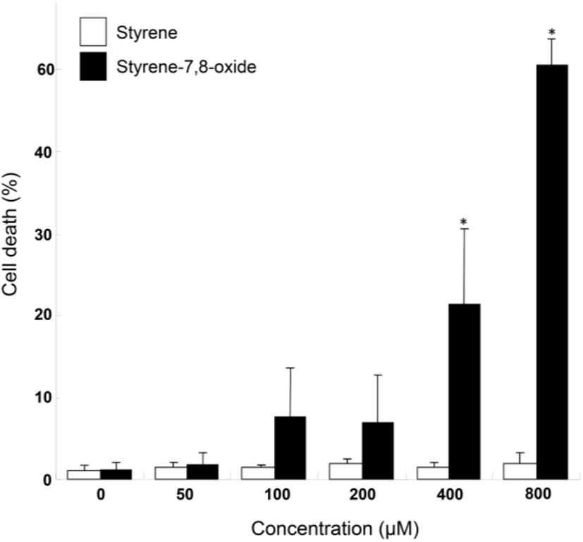
Although styrene cytotoxicity has been tested in several cell types (Cruzan et al., 2002; Corsi et al., 2007), including neuronal cells (Daré et al., 2004; Moujahed et al., 2020), a corresponding investigation has not been performed in testicular cells, particularly Leydig cells. In this study, we first monitored the nuclear fragmentation in Leydig cells after exposure to styrene or styrene-7,8-oxide. Nuclear fragmentation is a well-known morphological hallmark of apoptotic cells (Negri et al., 1996). Surprisingly, Hoechst 33258 cell staining showed that styrene exposure in TM3 Leydig cells did not exhibit nuclear fragmentation at any concentration, even at 800 μM, which is clearly pharmacological (Fig. 1A). In contrast, nuclear fragmentation was seen in styrene-7,8-oxide-exposed cells (Fig. 1B). However, nuclear fragmentation was only observed at higher concentrations (>400 μM) of styrene-7,8-oxide (Fig. 1B). Furthermore, cell death determined by sub-G1 analysis of the cell cycle confirmed that while styrene treatment did not induce significant cell death with any concentration (Fig. 2), styrene-7,8-oxide treatment showed significant cell death numerically at higher concentrations (Fig. 2). These results indicate that cytotoxicity-mediated cell death in Leydig cells is more susceptible to styrene-7,8-oxide than to styrene. Similar results were observed in Leydig cells after exposure to benzo[a]pyrene (B[a]P) or B(a)P diolepoxide (BPDE) (Chung et al., 2007). These differences in cell death susceptibility may occur because chemicals that retain oxide compounds are more likely able to form DNA adducts, causing DNA damage and cytotoxicity-mediated cell death i.e., apoptosis (Venkatachalam et al., 1993).
dr-26-3-99-g2
Fig. 2. Effects of styrene or styrene-7,8-oxide on on cell death in TM3 Leydig cells. Cell death after treatment with various concentrations of styrene (white rectangle) and styrene-7,8-oxide (black rectangle) (0–800 μM) for 48 h. Cell death was assessed by flow cytometry. The percentage of cells with a sub-G1 DNA content was taken as a measure of cell death. At least three independent experiments were performed and data shown are the mean±SD. * p<0.01 compared to control.
Download Original Figure

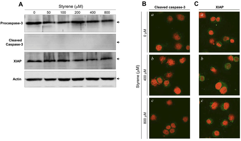
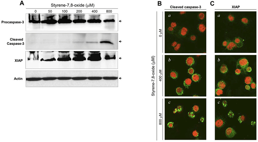
The activation of caspase-3 and upregulation of XIAP have been associated with the induction and suppression of apoptosis, respectively (Porter and Jänicke, 1999; Liston et al., 2003). Following styrene treatment, procaspase-3 and XIAP protein levels did not show significant changes, and cleaved (active) forms of caspase-3 were not detected (Fig. 3A). Consistent with the western blot results, the active forms of caspase-3 and XIAP proteins were not prominently altered in the cytoplasm of cells treated with styrene (Fig. 3B). These results confirmed that apoptosis does not occur after styrene exposure and implied that the cytotoxic effect of styrene on TM3 Leydig cells does not appear be fatal. In contrast to styrene, styrene-7,8-oxide induced cell death in an apoptotic fashion, as seen in caspase-3 activation and increased the expression of XIAP proteins (Fig. 4A). This finding was also confirmed by immunostaining for the active forms of caspase-3 and XIAP (Fig. 4B). These results suggest that TM3 Leydig cells retain an apoptotic mechanism, but the insufficiency or lack of cytochrome P450 enzymes (i.e., CYP 2E1) responsible for the conversion of styrene to styrene-7,8-oxide is the reason why TM3 Leydig cells could be protected from apoptosis induced by styrene exposure.
dr-26-3-99-g3
Fig. 3. Caspase-3 and Xiap proteins in TM3 Leydig cells after exposure to various concentrations of styrene. (A) Western blot analysis of caspase-3, cleaved (activated) caspase-3 and Xiap after styrene treatment for 48 h. Equal amounts of protein (30 μg) were separated by SDS-PAGE and immunoblotted using a cleaved form specific antibody for corresponding proteins. Actin expression was examined as a loading control. Representative confocal images of the activated caspase-3 (B) and XIAP (C). Cells were treated with styrene for 48 h. The cells were then cytospun, fixed, and immunostained with an active form specific antibody for corresponding proteins. Green fluorescence (FITC), target proteins; Red fluorescence (PI), nucleus. a, Control; b, 400 μM styrene-treated; c, 800 μM styrene-treated. Original magnification: ×800.
Download Original Figure
dr-26-3-99-g4
Fig. 4. Alterations of caspase-3 and Xiap proteins in TM3 Leydig cells after exposure to various concentrations of styrene-7,8-oxide. (A) Western blot analysis of caspase-3, cleaved (activated) caspase-3 and Xiap after styrene-7,8-oxide treatment for 48 h. Equal amounts of protein (30 μg) were separated by SDS-PAGE and immunoblotted using a cleaved form specific antibody for corresponding proteins. Actin expression was examined as a loading control. Immunolocalization of the activated caspase-3 (B) and XIAP (C). Cells were treated with styrene-7,8-oxide for 48 h. The cells were then cytospun, fixed, and immunostained with an active form specific antibody for corresponding proteins. Green fluorescence (FITC), target proteins; Red fluorescence (PI), nucleus. a, Control; b, 400 μM styrene-treated; c, 800 μM styrene-treated. Original magnification: ×800.
Download Original Figure

Taken together, the results obtained in this study demonstrate a fundamental idea that Leydig cells are capable of protecting themselves from cytotoxicity-mediated apoptosis as a result of styrene exposure in vitro. However, styrene exposure in vivo, in experimental animals, is thought to exhibit adverse effects on Leydig cells, because styrene can be metabolized to styrene-7,8-oxide in the liver. It remains unclear whether the steroid-producing function, i.e., steroidogenesis, of Leydig cells is also unaffected by exposure to styrene. We believe that this finding is particularly important now, since polystyrene is a major component of microplastics, which have received increasing attention as an environmental risk factor for human health. Therefore, further studies are needed to elucidate the endocrine disrupting potential of styrene in Leydig cells.

Conflict of interests

The authors declare no potential conflict of interest.

Acknowledgements

This work was supported by Korea Environment Industry & Technology Institute (KEITI) through Technology Development Project for Safety Management of Household Chemical Products, funded by Korea Ministry of Environment (MOE) (1485017593).

Authors’ contributions

Conceptualization: Chung JY, Yu WJ, Kim JM.

Data curation: Chung JY, Park JE.

Formal analysis: Kim YJ, Lee SJ

Methodology: Chung JY, Park JE.

Validation: Chung JY, Park JE, Kim JM.

Investigation: Chung JY, Park JE, Kim JM.

Writing-original draft: Chung JY, Park JE.

Writing-review & editing: Chung JY, Park JE, Kim YJ, Lee SJ, Yu WJ, Kim JM.

Ethics approval

This article does not require IRB/IACUC approval because there are no human and animal participants.

REFERENCES

1.

FM Abdel-MaksoudFAZ AliBT Akingbemi 2019; Prenatal exposures to bisphenol A and di (2-ethylhexyl) phthalate disrupted seminiferous tubular development in growing male rats. Reprod Toxicol. 88:85-90

2.

BT AkingbemiCM SottasAI KoulovaGR KlinefelterMP Hardy 2004; Inhibition of testicular steroidogenesis by the xenoestrogen bisphenol A is associated with reduced pituitary luteinizing hormone secretion and decreased steroidogenic enzyme gene expression in rat Leydig cells. Endocrinology. 145:592-603

3.

JY ChungJY KimYJ KimSJ JungJE ParkSG LeeJT KimS OhCJ LeeYD YoonYH YooJM Kim 2007; Cellular defense mechanisms against benzo[a]pyrene in testicular Leydig cells: Implications of p53, aryl-hydrocarbon receptor, and cytochrome P450 1A1 status. Endocrinology. 148:6134-6144

4.

JY ChungYJ KimJY KimSG LeeJE ParkWR KimYD YoonKS YooYH YooJM Kim 2011; Benzo[a]pyrene reduces testosterone production in rat Leydig cells via a direct disturbance of testicular steroidogenic machinery. Environ Health Perspect. 119:1569-1574

5.

P CorsiA D’AprileB NicoGL CostaG Assennato 2007; Synaptic contacts impaired by styrene-7,8-oxide toxicity. Toxicol Appl Pharmacol. 224:49-59

6.

G CruzanGP CarlsonKA JohnsonLS AndrewsMI BantonC BevanJR Cushman 2002; Styrene respiratory tract toxicity and mouse lung tumors are mediated by CYP2F-generated metabolites. Regul Toxicol Pharmacol. 35:308-319

7.

E DaréR TofighiL NuttMV VettoriM EmgårdA MuttiS Ceccatelli 2004; Styrene 7,8-oxide induces mitochondrial damage and oxidative stress in neurons. Toxicology. 201:125-132

8.

YY GongR KishiY KatakuraE TsukishimaK FujiwaraS KasaiT SatohF SataT Kawai 2002; Relation between colour vision loss and occupational styrene exposure level. Occup Environ Med. 59:824-829

9.

KV GopalC WuEJ MooreGW Gross 2011; Assessment of styrene oxide neurotoxicity using in vitro auditory cortex networks. ISRN Otolaryngol. 2011:204804

10.

FP GuengerichDH KimM Iwasaki 1991; Role of human cytochrome P-450 IIE1 in the oxidation of many low molecular weight cancer suspects. Chem Res Toxicol. 4:168-179

11.

P ListonWG FongRG Korneluk 2003; The inhibitors of apoptosis: There is more to life than Bcl2. Oncogene. 22:8568-8580

12.

C MöllerL OdkvistB LarsbyR ThamT LedinL Bergholtz 1990; Otoneurological findings in workers exposed to styrene. Scand J Work Environ Health. 16:189-194

13.

S MoujahedA RuizD HallegueM Sakly 2022; Quercetin alleviates styrene oxide-induced cytotoxicity in cortical neurons in vitro via modulation of oxidative stress and apoptosis. Drug Chem Toxicol. 45:1634-1643.

14.

C NegriR BernardiM DonzelliE ProsperiAI Scovassi 1996; Sequence of events leading to apoptosis in long term cultured HeLa cells. Cell Death Differ. 3:425-430.

15.

AG PorterRU Jänicke 1999; Emerging roles of caspase-3 in apoptosis. Cell Death Differ. 6:99-104

16.

M Sliwinska-KowalskaA FuenteE Zamyslowska-Szmytke 2020; Cochlear dysfunction is associated with styrene exposure in humans. PLoS One. 5:e0227978

17.

K TraoreDB Martinez-ArguellesV PapadopoulosH ChenBR Zirkin 2016; Repeated exposures of the male Sprague Dawley rat reproductive tract to environmental toxicants: Do earlier exposures to di-(2-ethylhexyl)phthalate (DEHP) alter the effects of later exposures. Reprod Toxicol. 61:136-141

18.

S VenkatachalamMF DenissenkoN AlviAA Wani 1993; Rapid activation of apoptosis in human promyelocytic leukemic cells by (+/–)-anti-benzo[a]pyrene diol epoxide induced DNA damage. Biochem Biophys Res Commun. 197:722-729

19.

BR Zirkin 1998; Spermatogenesis: Its regulation by testosterone and FSH. Semin Cell Dev Biol. 9:417-42