Development & Reproduction
Korean Society of Developmental Biology
Research article

Signal Transduction of Constitutive Activating and Inactivating Eel Lutropin/Choriogonadotropin Receptor (Eel LH/CGR) Mutants by Recombinant Equine Chorionic Gonadotropin (Rec-eCG)

Kwan-Sik Min1,2,3,https://orcid.org/0000-0002-5451-3085, Sei Hyen Park1https://orcid.org/0009-0009-7670-0090, Ha-Rin Jeong2https://orcid.org/0009-0002-1322-4938, Jae-Hyek Park2https://orcid.org/0009-0000-8650-7184, Munkhzaya Byambaragchaa3https://orcid.org/0000-0002-0277-1816
1Graduate School of Animal BioSciences, Hankyong National University, Anseong 17579, Korea
2Division of Animal BioScience, School of Animal Life Convergence Sciences, Hankyong National University, Anseong 17579, Korea
3Carbon-Neutral Resources Research Center, Institute of Genetic Engineering, Hankyong National University, Anseong 17579, Korea
Corresponding author Kwan-Sik Min, Division of Animal BioScience, School of Animal Life Convergence Sciences, Hankyong National University, Anseong 17579, Korea. Tel: +82-31-670-5421, E-mail: ksmin@hknu.ac.kr

© Copyright 2024 The Korean Society of Developmental Biology. This is an Open-Access article distributed under the terms of the Creative Commons Attribution Non-Commercial License (http://creativecommons.org/licenses/by-nc/4.0/) which permits unrestricted non-commercial use, distribution, and reproduction in any medium, provided the original work is properly cited.

Received: Sep 16, 2024 ; Revised: Nov 08, 2024 ; Accepted: Nov 19, 2024

Published Online: Dec 31, 2024

Abstract

Lutropin/choriogonadotropin receptor (LH/CGR) is a member of the G protein-coupled receptor superfamily. LH/CGRs in fish and mammalian species have been reported to contain naturally occurring, constitutively activating, and inactivating mutations in highly conserved regions. The present study was designed to determine the functional aspect of eel LH/CGR signal transduction. Biochemical analysis was performed using cells transfected with wild-type eel LH/CG (eel LH/CGR-wt) or with activating (designated eel LH/CGR-M410T, L469R, and D590Y) and inactivating (eel LH/CGR-D 417N and Y558F) mutants. We also generated a mutant (eel LH/CGR-t651) in which the C-terminal cytoplasmic tail was truncated at residue 651. Activating mutant cells expressing eel LH/CGR-M410T, L469R, and D590Y exhibited 1.4-, 8.7-, and 4.0-fold increases in the basal cAMP response, respectively, without recombinant equine chorionic gonadotropin (rec-eCG) agonist treatment. In inactivating mutants (eel LH/CGR-D417N and Y558F), the cyclic adenosine monophosphate (cAMP) response did not result in completely impaired signal transduction. However, the eel LH/CGR-t651 mutant did not exhibit any cAMP signaling following high-agonist treatment. Rmax values did not increase with further rec-eCG agonist stimulation. Our results suggest that constitutively activating and inactivating eel LH/CGR mutants with highly conserved amino acids exhibit a significant signal transduction pathway for glycoprotein hormone receptors. Eel LH/CGRs in activating and inactivating mutants are usually processed by receptor-mediated signaling following rec-eCG agonist stimulation.

Keywords: Eel lutropin/choriogonadotropin receptor (eel LH/CGR); Constitutive activation; Inactivation; cyclic adenosine monophosphate (cAMP) response; Recombinant equine chorionic gonadotropin (rec-eCG)

INTRODUCTION

G protein-coupled receptors (GPCRs) form the largest class of transmembrane cell surface receptors and regulate diverse intracellular signaling pathways in response to a wide range of extracellular stimuli (Lefkowitz & Shenoy, 2005; Lefkowitz et al., 2023). Gonadotropin receptors such as lutropin/choriogonadotropin receptors (LH/CGR) belong to a large superfamily of GPCRs with seven transmembrane-spanning domains and large extracellular and intracellular regions (Choi et al., 2023).

LH/CGRs mediate intracellular effects through G protein activation (α, β, and γ) (Min et al, 2019, 2020). These Gα subunits are characterized into four classes: Gαs, Gαi, Gαq, and Gα12 (Syrovatkina et al., 2016). Gαs and Gαi stimulate and inhibit adenylate cyclase, respectively, thereby regulating cyclic AMP responsiveness. LH/CGR is involved in the receptor-mediated cyclic adenosine monophosphate (cAMP) response (Tao et al., 2000; Bhaskaran & Ascoli, 2005; Byambaragchaa et al., 2021a) and the phosphorylation and activation of extracellular-related kinase (pERK1/2) (Martinelle et al., 2004; Tao, 2006; Shiraishi & Ascoli, 2007).

Equine choriogonadotropin (eCG) is a glycoprotein hormone secreted by trophoblastic cells into the bloodstream of pregnant mares during the 2nd to 5th months of pregnancy, with a peak level at 70–80 days of gestation (Min et al., 1996, 2004; Rodríguez et al., 2023). eCG consists of a common α-subunit and a hormone specific β-subunit (Min et al., 2019, 2020; Lee et al., 2021). A single gene encodes both the CG β-subunit and LH β-subunit in horses (Sherman et al., 1992; Min et al., 1996, 2004), and results in follicle-stimulating hormone (FSH)-like and luteinizing hormone (LH)-like response in non-equid species (Lee et al., 2021).

In previous studies, LH/CGR mutations have been reported, where activating mutants could trigger precocious puberty in boys, but inactivating mutants could cause anovulation in women and blocked sexual maturation in men (Thermmen & Huhtaniemi, 2000). These mutants were shown to be constitutively active in the absence of agonist stimulation, with basal cAMP level elevated up to several fold compared to the wild-type receptor (Shenker et al., 1993; Zhang et al., 2005, 2007; Galet & Ascoli, 2006; Latronico & Segaloff, 2007). Recently, we reported the structural and functional properties of equine LH/CGR activating and inactivating mutants (Byambaragchaa et al., 2021a) and equine follicle stimulating hormone receptor (FSHR; Byambaragchaa et al., 2022a).

Most GPCRs have several sites in their C-terminal regions that undergo modification and activate downstream signaling in response to agonist-receptor binding. These C-terminal modifications include phosphorylation (serine and threonine), highly conserved palmitoylation (two cysteines), and tri-leucine residues.

We reported that the C-terminal phosphorylation sites play a pivotal role in the signal transduction of equine FSHR (Seong et al., 2020) and equine LH/CGR (Byambaragchaa et al., 2022b). In eel LH/CGR and FSHR, we reported that activating mutants exhibited a large increase in the basal cAMP response without agonist stimulation (Byambaragchaa et al., 2021b). A C-terminal deletion mutant of eel FSHR led to a decrease in the maximal cAMP response (Kim et al., 2018).

In the present study, we demonstrated that constitutively activating mutants in eel LH/CGR exhibited a high fold increase in basal cAMP response, but inactivating mutants showed a low cAMP response under high concentrations of the recombinant equine chorionic gonadotropin (rec-eCG) agonist.

MATERIALS AND METHODS

1. Materials

Oligonucleotides were synthesized, and mutated DNAs was sequenced by Genotech (Daejeon, Korea). The pGEM-T Easy Cloning Vector was purchased from Promega (Madison, WI, USA). DNA ligation reagents, endonucleases, PCR reagents, and restriction enzymes were purchased from Takara Bio (Shiga, Japan). Mammalian expression vector pCORON1000 SP VSV-G was purchased from Amersham Biosciences (Piscataway, NJ, USA). The pcDNA3 mammalian expression vector, CHO-S cells, FreeStyle MAX reagent, and FreeStyle CHO expression medium were purchased from Invitrogen (Carlsbad, CA, USA). CHO-K1 cells were obtained from the Korean Cell Line Bank (Seoul, Korea). Ham’s F-12 medium, Opti-MEM I, and serum-free CHO-S-SFM II were purchased from Gibco (Grand Island, NY, USA). The cAMP homogeneous time-resolved fluorescence (HTRF) assay kit was purchased from Cisbio (Codolet, France). The QIAprep-Spin plasmid kit was purchased from Qiagen (Hilden, Germany) and disposable flasks were purchased from Corning (Corning, NY, USA). CentriPlus centrifugal filters were purchased from Amicon Bio Separation (Billerica, MA, USA). The PMSG enzyme-linked immunosorbent assay (ELISA) kit was purchased from DRG International (Mountainside, NJ, USA). All other reagents were purchased from Sigma-Aldrich (St. Louis, MO, USA).

2. Site-directed mutagenesis and vector construction of eel LH/CGRs

To construct point mutations in eel LH/CGR activating and inactivating mutants, we used cDNA encoding full-length eel LH/CGR-wt using an overlap extension PCR strategy to substitute specific amino acids, as described previously (Byambaragchaa et al., 2021b). The PCR products were ligated into the pGEMT-Easy vector and the sequence of each mutant was confirmed by DNA sequencing. Full-length fragments were cleaved using the XhoI and EcoRI restriction enzymes and ligated into the pCORON SP VSVG mammalian expression vector.

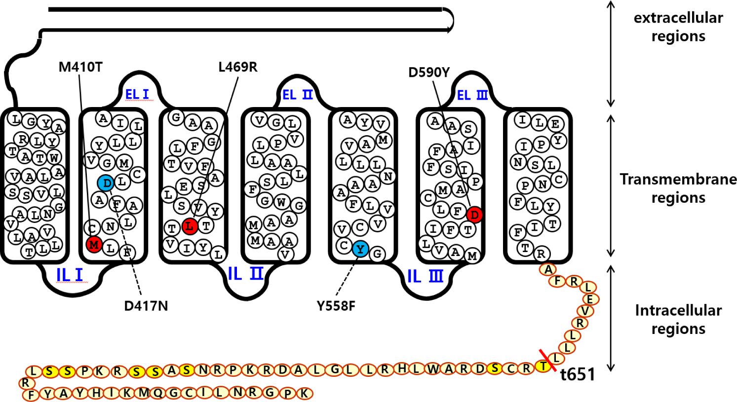
The signal sequence region was removed from eel LH/CGR and the fragments were inserted under the VSVG-tag signal sequence of the mammalian expression vector. Finally, the direction of insertion was confirmed by restriction enzyme digestion. We constructed three activating mutants (eel LH/CGR-M410T, L469R, and D590Y), two inactivating mutants (eel LH/CGR-D417N and Y558F) in the previous studies (Byambaragchaa et al., 2021b), and one deletion mutant (eel LH/CGR-t641) at the C-terminal cytoplasmic tail region. Fig. 1 shows a schematic diagram of eel LH/CGR-wt and the mutants, denoting the amino acids of the conserved activation and inactivation sites in eel LH/CGR.
dr-28-4-141-g1
Fig. 1. Schematic representation of the structure of eel lutropin/choriogonadotropin receptor (eel LH/CGR)-wt and mutants. The amino acid sequences of the transmembrane regions I–IIV and the C-terminal region presuming potential phosphorylation sites were shown. The locations of the constitutively activating mutations (M410T, K469R, and D590Y) and two inactivating mutations (D417N and Y558F) are indicated. The red circle indicates the constitutively activating mutations, and the sky-blue circles indicate inactivating mutations. The C terminal region mutant had deletion of the seven phosphorylation sites. The 7 potential phosphorylation sites (serine and threonine residues) are shown. The truncation site is present at the 651st amino acid. Yellow colors indicate the potential phosphorylation sites.
Download Original Figure
3. Transient transfection into CHO-K1

Plasmids cloned into the pCORON SP VSVG expression vector were transfected into CHO-K1 cells using liposome-mediated transfection, as previously reported (Byambaragchaa et al., 2024). The cells were grown to 80%–90% confluence in 6-well plates. Plasmid DNA and transfection reagent were mixed with Opti-MEM and incubated for 20 min at room temperature. The cells were washed with Opti-MEM and the DNA-Lipofectamine complex was added. Fresh growth medium containing 20% FBS was added to each well 5 h after transfection. The cells were subjected to cAMP analysis at 48 h post-transfection.

4. Production of rec-eCG in CHO suspension cells

The pcDNA3 mammalian vectors were transfected into CHO suspension (CHO-S) cells using the FreeStyle MAX reagent, as reported in a previous study (Lee et al., 2021). Briefly, cells were cultured in 30 mL of CHO expression medium for 3 days. The cells were passaged at a 5–6×10⁵ cells/mL in 125 mL CHO expression medium in a disposable spinner flask. The cell density was approximately 1.2–1.5 cells/mL on the day of transfection. Plasmid DNA (160 μg) and FreeStyle MAX reagent (160 μL) were mixed gently with 1.2 mL of OptiPRO serum-free medium and media samples were mixed and incubated for 5 min at RT. The mixed samples (2.4 mL) were gently added to the cell suspension flask. Finally, for the rec-eCG protein assay, the supernatant was collected on day nine post-transfection. The collected media were concentrated by CentriPlus centrifugal filters and quantified by PMSG ELISA with an anti-PMSG monoclonal antibody, horseradish peroxidase (HRP)-conjugated antibody, and TMB substrate, following the manufacturer’s instructions in the previous study (Lee et al., 2021).

5. Analysis of cAMP levels via homogenous time-resolved fluorescence assays

cAMP accumulation in CHO-K1 cells expressing eel LH/CGR-wt and mutants was measured using a cAMP Dynamics 2 competitive immunoassay kit (Cisbio Bioassays, Codolet, France), as described in a previous study (Byambaragchaa et al., 2024). Briefly, transfected cells were diluted in 0.5 mM IBMX to inhibit cAMP degradation and seeded in 384-well plates (10,000 cells per well). Rec-eCG ligand (5 μL) was added to each well, and the cells were stimulated at RT for 30 min. The cAMP response was analyzed by measuring the decrease in HTRF energy transfer (665 nm/620 nm) using a TriStar2 S LB942 mi microplate reader (BERTHOLD Tech, Wildbad, Germany). The results were calculated from the ratio of fluorescence at 665 and 620 nm and expressed as Delta F% (cAMP inhibition) according to the following equation: Delta F% = (standard or sample ratio − mock transfection) 100 / mock transfection.

The cAMP concentration for Delta F% values was calculated using GraphPad Prism software (version 6.0; GraphPad Software, La Jolla, CA, USA).

6. Data analysis

The Multalin multiple sequence alignment tool was used for the sequence analysis. Dose-response curves were generated for experiments performed in duplicates. GraphPad Prism 6.0 (San Diego, CA, USA) was used to analyze the cAMP response, EC50 values, and stimulation curves. Curves fitted in a single experiment were normalized to background signals measured in mock-transfected cells. Each curve was plotted using the data from three independent experiments. Data were analyzed by one-way analysis of variance (ANOVA), followed by Tukey’s comparison test using GraphPad Prism v.6.0, as indicated in the figure captions. Statistical significance was determined at the following levels: * p<0.05 and ** p<0.01, indicating a significant difference between groups.

RESULTS

1. Biological activities of the constitutively activating mutants of eel LH/CGR

The in vitro biological activity was assessed using transfected CHO-K1 cells expressing eel LH/CGR-wt and activating mutants. The results of cAMP activation are shown in Fig. 2 and Table 1. The basal and Rmax cAMP responses in eLH/CGR-wt were 3.8 and 64.5 nM/104 cells, respectively (Table 1). cAMP responsiveness increased in a dose-dependent manner. The half-maximal effective concentration (EC50) value of rec-eCG-stimulated cAMP was approximately 58.66 ng/mL. The dose-response curve of the eLH/CGR-activating mutants was slightly shifted to the left compared with that of eLH/CGR-wt. Specifically, the basal cAMP response was very high compared to that observed in eLH/CGR-wt. The activating mutants (L469R and D590Y) showed 8.7- and 4.0-fold increases in the basal cAMP response, respectively, without rec-eCG agonist treatment. However, the M410T mutant did not show any difference in the basal cAMP response (Fig. 3).

dr-28-4-141-g2
Fig. 2. Total cAMP levels stimulated by recombinant-eCG (rec-eCG) in CHO-K1 cells transfected with eel LH/CGR-wt and constitutively activating eLH/CGR. CHO-K1 cells transiently transfected with eLH/CGR-wt and mutants were seeded in 384-well plates (10,000 cells/well) at 24 h post-transfection. The cells were stimulated with the rec-eCG proteins for 30 min at room temperature. cAMP production was detected using a homogeneous time-resolved fluorescence assay and presented as Delta F%. cAMP concentrations were calculated using GraphPad Prism software. Each data point represents mean±SD of the mean of triplicate experiments. The mean data was fitted to an equation to derive a single-phase exponential decay curve. LH, luteinizing hormone; CGR, choriogonadotropin receptor; cAMP, cyclic adenosine monophosphate.
Download Original Figure
Table 1. Bioactivities of constitutive activating eel LH/CGR mutants
Eel LH/CG receptors cAMP responses
Basal1 (nM/104 cells) Log (EC50) (ng/mL) Rmax2 (nM/104 cells)
Eel LH/CGR-wt 3.8±1.0 58.66 (48.1 to 75.3)3 64.5±1.7
Eel LH/CGR-M410T 5.2±0.9 (1.4-fold) 21.7 (16.8 to 30.8) 42.4±1.2
Eel LH/CGR-L469R 33.2±0.7 (8.7-fold) 708 (373.4 to 6,965) 67.9±3.4
Eel LH/CGR-D590Y 15.3±1.5 (4.0-fold) 31.1 (22.5 to 50.2) 64.8±2.2

Values are the means±SEM of triplicate experiments. The log (EC50) values were determined from the concentration-response curves obtained from the in vitro bioassays.

Basal cAMP level average without agonist treatment.

Rmax average cAMP level/104 cells.

Geometric mean (95% confidence limit) of at least three experiments.

LH, luteinizing hormone; CGR, choriogonadotropin receptor; cAMP, cyclic adenosine monophosphate; EC50, effective concentration.

Download Excel Table
dr-28-4-141-g3
Fig. 3. The results of basal cAMP and Rmax level responsiveness in activating mutants. The basal and Rmax level responses presented in Fig. 2 are displayed using a bar graph. The results are expressed as mean±SD of a single representative experiment performed in triplicate. * Statistically significant differences were calculated by a one-way ANOVA followed by Tukey’s comparison test (* p<0.05) and (** p<0.01). cAMP, cyclic adenosine monophosphate.
Download Original Figure

The L469R mutant showed the highest basal cAMP response, corresponding to approximately 50% of the maximal value observed in eLH/CGR-wt (Table 1). However, cAMP activity in the L468R mutant did not constitutively increase despite the high concentration of the rec-eCG agonist (Fig. 2). In the M410T mutant, the Rmax level was the lowest among all activating mutants. EC50 values in the M410T and D590Y mutants showed approximately 2-fold increase, but that in the L469R mutant could not be calculated exactly because of the high basal cAMP level priot to treatmemnt with rec-eCG agonist (100 ng/mL) (Table 1). Rmax values for L469R and D590Y mutants were almost similar to that of eel LH/CGR-wt, but the value for M410T mutant was significantly lower (42.4 nM/104 cells), which is approximately 65% of the Rmax value of the eel LH/CGR-wt (Table 1).

2. Biological activities of inactivating mutants of eel LH/CGR

Eel LH/CGRs with inactivating mutations-D417N and Y558F were evaluated by quantifying cAMP accumulation in cells incubated with increasing concentrations of rec-eCG. The basal cAMP activity was not affected, as shown in Figs. 4 and 5 and Table 2. However, cAMP signaling was not completely impaired in these mutants, despite lower activation (Fig. 4). These Rmax values were approximately 50% of the corresponding values for eel LH/CGR-wt (Table 2). The EC50 value was approximately 108 ng/mL. However, the EC50 values of D417N and Y558F decreased 2.3- and 3.2-fold, respectively, demonstrating that detected approximately 253.9 ng/mL and 345 ng/mL, respectively.

dr-28-4-141-g4
Fig. 4. Total cAMP levels stimulated by recombinant-eCG (rec-eCG) in CHO-K1 cells transfected with eel LH/CGR-wt and inactivating mutants (eel LH/CGR-D417 and Y558F) and eel LH/CGR-t641. The cells were stimulated with the rec-eCG proteins for 30 min at room temperature. cAMP concentrations were calculated using GraphPad Prism. Each data point represents mean±SD of the mean of triplicate experiments. The mean data were fitted to an equation to derive a single-phase exponential decay curve. LH, luteinizing hormone; CGR, choriogonadotropin receptor; cAMP, cyclic adenosine monophosphate.
Download Original Figure
dr-28-4-141-g5
Fig. 5. The results of basal cAMP and Rmax level responsiveness in inactivating mutants and C-terminal deleted mutant. The results are expressed as mean±SD of a single representative experiment performed in triplicate. * Statistically significant differences were calculated by a one-way ANOVA followed by Tukey’s comparison test (** p<0.01). Representative data are shown, and the graphs contain the mean±SE values of three independent experiments. cAMP, cyclic adenosine monophosphate.
Download Original Figure
Table 2. Bioactivities of inactivating eel LH/CGR mutants and C-terminal deleted mutant
Eel LH/CG receptors cAMP responses
Basal1 (nM/104 cells) Log (EC50) (ng/mL) Rmax2 (nM/104 cells)
Eel LH/CGR-wt 2.2±1.4 108 (86.7 to 144.5)3 76.4±2.0
Eel LH/CGR-D417N 0.8±0.3 253.9 (2.3-fold) (227 to 288.2) 44.2±0.7
Eel LH/CGR-Y558F 1.6±0.5 345.4 (3.2-fold) (12.4 to 16.7) 38.8±1.1
Eel LH/CGR-t651 0.7±0.02 - -

Values are the means±SEM of triplicate experiments. The log (EC50) values were determined from the concentration-response curves obtained from the in vitro bioassays.

Basal cAMP level average without agonist treatment.

Rmax average cAMP level/104 cells.

Geometric mean (95% confidence limit) of at least three experiments.

LH, luteinizing hormone; CGR, choriogonadotropin receptor; cAMP, cyclic adenosine monophosphate; EC50, effective concentration.

Download Excel Table
3. Biological activity of eel LH/CGR-t651 mutant

The eel LH/CGR also has a long C-terminal region containing approximately seven phosphorylation sites, as shown in Fig. 1. To assess the function of potential phosphorylation sites in cAMP signal transduction, eel LH/CGR-wt was truncated at amino acid 651 of the C-terminal region and the truncated mutant was referred to as eLH/CGR-t651. This mutation resulted in completely impaired cAMP responsiveness, as shown in Figs. 4 and 5 and Table 2. Thus, the Rmax value did not show any specific variation, indicating that the phosphorylation regions in the eel LH/CGR C-terminal region played a pivotal role in the PKA signal transduction pathway.

DISCUSSION

Based on the results of the present study, our findings clearly show that activating mutants exhibited constitutively active cAMP signaling (M410T, L460R, and D590Y), while inactivating (D417N and Y558F) mutants exhibited highly decreased and C-terminus deleted mutant (eel LH/CGR-t651) exhibited completely impaired cAMP responsiveness. However, all receptor mutants showed effective response to the rec-eCG agonist treatment. These highly conserved sites in the transmembrane region are important for downstream signal transduction. Therefore, we concluded that activating eel LH/CGR mutants exhibit continuous signaling without agonist stimulation. Phosphorylation sites in the C-terminal region of eel LH/CGR are essential for rec-eCG-induced phosphorylation.

Recently, we characterized eel LH/CGRs by rec-eel LH stimulation at the same sites, and found that activating mutants displayed 4–19-fold increase in basal cAMP responsiveness without agonist treatment (Byambaragchaa et al., 2021b). In equine LH/CGR, three constitutively activating mutants (L457R, D564G, and D578Y) exhibited 16.9-, 16.4-, and 11.2-fold increases in the basal cAMP response, respectively (Byambaragchaa et al., 2021a). Cells expressing eel LH/CGR-D576G mutant, which contains a highly conserved aspartic acid, exhibited a 5.8-fold increase following rec-eel LH and rec-eCG stimulation (Choi et al., 2023). Basal cAMP activity exhibited a 4.5-fold increase in COS-7 cells in hLH/CGR-D578G (Shenker et al., 1993) and hLH/CGR-L457R (Zhang et al., 2005, 2007; Galet & Ascoli, 2006; Latronico & Segaloff, 2007). In the present study, our data on the activation of eel LH/CGR-L469R and D590Y mutants were concordant with previous reports showing that the basal cAMP response resulted in a high increase without agonist stimulation.

In the inactivating mutants, many studies have reported in equine LH/CGR-D405N, -R464H, and Y546F (Byambaragchaa et al., 2021a), hLH/CGR-V144F (Richter-Unruh et al., 2005), hLH/CGR-F194V (Gromoll et al., 2002), rat LH/CGR-D383N, -R442H, and -Y524F (Dhanwada et al., 1996; Min et al., 1998), indicating that any mutants were completely impaired or not impaired. However, inactivating mutants did not affect the basal cAMP response.

Interestingly, our results in the presented study are no activity, in contrast to previously reported data for equine LH/CGR-t656 (Byambaragchaa et al., 2022b), equine FSHR-t641 (Seong et al., 2020), and eel FSHR-t614 (Kim et al., 2018), showing that agonist-induced receptors are not completely impaired in the cAMP response. In the present study, eel LH/CGR-t651 mutant were completely impaired. Thus, we suggest that the C-terminal regions of eel LH/CGR are necessary for agonist-induced uncoupling, with only seven phosphorylation sites. Thus, phosphorylation-dependent structure of the GPCR C-terminal region has been reported to modulate arrestin binding, and therefore, signaling is modulated in arrestin-dependent pathways (Guillien et al., 2023). Although this mechanism is not well understood, phosphorylation-and β-arrestin-dependent pathways may exist in truncated mutants for the internalization of eel LH/CGR. Thus, we suggest that conformational changes in the serine and threonine residues of the C-terminal domain of GPCR play an important role in the downstream signaling pathway.

In conclusion, our study showed that the activating mutants greatly increased the basal cAMP response, whereas the inactivating mutants decreased the Rmax cAMP response and EC50 value.

C-terminal deletion of potential phosphorylation sites completely impaired the cAMP response. The results of the present study suggest that there is a strong correlation between highly conserved activating and inactivating sites. Although not revealed in this study, the highly conserved regions in the C-terminus of eel LH/CGR represent specific signaling pathways such as internalization. Taken together, PKA signaling via LH/CGR-mediated signaling must be further studied to determine whether any pathway involving the phosphorylation of downstream effectors of the MAPK pathway exists.

Conflict of interests

The authors declare no potential conflict of interest.

Acknowledgements

This study was funded by the Korean Research Foundation Program (grant number: RS-2024-00335122).

Authors’ contributions

Conceptualization: Min KS.

Data curation: Jeong HR, Park JH.

Formal analysis: Park SH.

Methodology: Byambaragchaa M.

Software: Jeong HR, Park JH.

Investigation: Byambaragchaa M.

Writing-original draft: Byambaragchaa M.

Writing-review & editing: Min KS, Park SH, Jeong HR, Park JH, Byambaragchaa M.

Ethics approval

This article does not require IRB/IACUC approval because there are no human and animal participants.

REFERENCES

1.

Bhaskaran RS, Ascoli M. 2005; The post-endocytotic fate of the gonadotropin receptors is an important determinant of the desensitization of gonadotropin responses. J Mol Endocrinol. 34:447-457

2.

Byambaragchaa M, Ahn TY, Choi SH, Kang MH, Min KS. 2022a; Functional characterization of naturally-occurring constitutively activating/inactivating mutations in equine follicle-stimulating hormone receptor. Anim Biosci. 35:399-409

3.

Byambaragchaa M, Choi SH, Kim DW, Min KS. 2021b; Constitutive activating eel luteinizing hormone receptor induce constitutively signal transduction and inactivating mutants impair biological activity. Dev Reprod. 25:133-143

4.

Byambaragchaa M, Joo HE, Kim SG, Kim YJ, Park GE, Min KS. 2022b; Signal transduction of C-terminal phosphorylation regions for equine luteinizing hormone/chorionic gonadotropin receptor (eLH/CGR). Dev Reprod. 26:1-12

5.

Byambaragchaa M, Park SH, Kim SG, Shin MG, Kim SK, Park MH, Kang MH, Min KS. 2024; Stable production of a recombinant single-chain eel follicle-stimulating hormone analog in CHO DG44 cells. Int J Mol Sci. 25:7282

6.

Byambaragchaa M, Seong HK, Choi SH, Kim DJ, Kang MH, Min KS. 2021a; Constitutively activating mutants of equine LH/CGR constitutively induce signal transduction and inactivating mutations impair biological activity and cell-surface receptor loss in vitro. Int J Mol Sci. 22:10723

7.

Choi SH, Byambaragchaa M, Kim DJ, Lee JH, Kang MH, Min KS. 2023; Specific signal transduction of constitutively activating (D576G) and inactivating (R476H) mutants of agonist-stimulated luteinizing hormone receptor in eel. Int J Mol Sci. 24:9133

8.

Dhanwada KR, Vijapurkar U, Ascoli M. 1996; Two mutations of the lutropin/choriogonadotropin receptor that impair signal transduction also interfere with receptor-mediated endocytosis. Mol Endocrinol. 10:544-554

9.

Galet C, Ascoli M. 2006; A constitutively active mutant of the human lutropin receptor (hLHR-L457R) escapes lysosomal targeting and degradation. Mol Endocrinol. 20:2931-2945

10.

Gromoll J, Schulz A, Borta H, Gudermann T, Teerds KJ, Greschniok A, Nieschlag E, Seif FJ. 2002; Homozygous mutation within the conserved Ala-Phe-Asn-Glu-Thr motif of exon 7 of the LH receptor causes male pseudohermaphroditism. Eur J Endocrinol. 147:597-608

11.

Guillien M, Mouhand A, Sagar A, Fournet A, Allemand F, Pereira GAN, Thureau A, Bernadó P, Banères J, Sibille N. 2023; Phosphorylation motif dictates GPCR c-terminal domain conformation and arrestin interaction. Structure. 31:1394-1406.e7

12.

Kim JM, Byambaragchaa M, Kang MH, Min KS. 2018; The C-terminal phosphorylation sites of eel follicle-stimulating hormone receptor are important role in the signal transduction. Dev Reprod. 22:143-153

13.

Latronico AC, Segaloff DL. 2007; Insights learned from L457(3.43)R, an activating mutant of the human lutropin receptor. Mol Cell Endocrinol. 260-262:287-293

14.

Lee SY, Byambaragchaa M, Choi SH, Kang HJ, Kang MH, Min KS. 2021; Roles of N-linked and O-linked glycosylation sites in the activity of equine chorionic gonadotropin in cells expressing rat luteinizing hormone/chorionic gonadotropin receptor and follicle-stimulating hormone receptor. BMC Biotechnol. 21:52

15.

Lefkowitz RJ, Rockman HA, Shim PJ, Liu S, Ahn S, Pani B, Rajagopal S, Shenoy SK, Bouvier M, Benovic JL, Liggett SB, Ruffolo RR, Bristow MR, Packer M. 2023; How carvedilol does not activate β2-adrenoceptors. Nat Commun. 14:7866

16.

Lefkowitz RJ, Shenoy SK. 2005; Transduction of receptor signals by β-arrestins. Science. 308:512-517

17.

Martinelle N, Holst M, Söder O, Svechnikov K. 2004; Extracellular signal-regulated kinases are involved in the acute activation of steroidogenesis in immature rat Leydig cells by human chorionic gonadotropin. Endocrinology. 145:4629-4634

18.

Min KS, Hattori N, Aikawa JI, Shiota K, Ogawa T. 1996; Site-directed mutagenesis of recombinant equine chorionic gonadotropin/luteinizing hormone: Differential role of oligosaccharides in luteinizing hormone- and follicle-stimulating hormone-like activities. Endocr J. 43:585-593

19.

Min KS, Hiyama T, Seong HH, Hattori N, Tanaka S, Shiota K. 2004; Biological activities of tethered equine chorionic gonadotropin (eCG) and its deglycosylated mutants. J Reprod Dev. 50:297-304

20.

Min KS, Liu X, Fabritz J, Jaquette J, Abell AN, Ascoli M. 1998; Mutations that induce constitutive activation and mutations that impair signal transduction modulate the basal and/or agonist-stimulated internalization of the Lutropin/Choriogonadotropin receptor. J Biol Chem. 273:34911-34919

21.

Min KS, Park JJ, Byambaragchaa M, Kang MH. 2019; Characterization of tethered equine chorionic gonadotropin and its deglycosylated mutants by ovulation stimulation in mice. BMC Biotechnol. 19:60

22.

Min KS, Park JJ, Lee SY, Byambragchaa M, Kang MH. 2020; Comparative gene expression profiling of mouse ovaries upon stimulation with natural equine chorionic gonadotropin (N-eCG) and tethered recombinant-eCG (R-eCG). BMC Biotechnol. 20:59

23.

Richter-Unruh A, Korsch E, Hiort O, Holterhus PM, Themmen AP, Wudy SA. 2005; Novel insertion frameshift mutation of the LH receptor gene: Problematic clinical distinction of Leydig cell hypoplasia from enzyme defects primarily affecting testosterone biosynthesis. Eur J Endocrinol. 152:255-259

24.

Rodríguez MC, Mussio PE, Villarraza J, Tardivo MB, Antuña S, Fontana D, Ceaglio N, Prieto C. 2023; Physiochemical characterization of a recombinant eCG and comparative studies with PMSG commercial preparations. Protein J. 42:24-36

25.

Shenker A, Laue L, Kosugi S, Merendino JJ, Minegishi T, Cutler GB. 1993; A constitutively activating mutation of the luteinizing hormone receptor in familial male precocious puberty. Nature. 365:652-654

26.

Sherman GB, Wolfe MW, Farmerie TA, Clay CM, Threadgill DS, Sharp DC, Nilson JH. 1992; A single gene encodes the β-subunits of equine luteinizing hormone and chorionic gonadotropin. Mol Endocrinol. 6:951-959

27.

Shiraishi K, Ascoli M. 2007; Lutropin/choriogonadotropin stimulate the proliferation of primary cultures of rat Leydig cells through a pathway that involves activation of the extracellularly regulated kinase 1/2 cascade. Endocrinology. 148:3214-3225

28.

Seong HK, Choi SH, Byambaragchaa M, Min KS. 2020; Signal transduction of C-terminal phosphorylation sites for equine follicle stimulating hormone receptor (eFSHR). J Anim Reprod Biotechnol. 35:155-162

29.

Syrovatkina V, Alegre KO, Dey R, Huang XY. 2016; Regulation, signaling, and physiological functions of G-proteins. J Mol Biol. 428:3850-3868

30.

Tao YX. 2006; Inactivating mutations of G protein-coupled receptors and diseases: Structure-function insights and therapeutic implications. Pharmacol Ther. 111:949-973

31.

Tao YX, Abell AN, Liu X, Nakamura K, Segaloff DL. 2000; Constitutive activation of G protein-coupled receptors as a result of selective substitution of a conserved leucine residue in transmembrane helix III. Mol Endocrinol. 14:1272-1282

32.

Thermmen APN, Huhtaniemi IT. 2000; Mutation of gonadotropins and gonadotropin receptors: Elucidating the physiology and pathophysiology of pituitary-gonadal function. Endocr Rev. 21:551-583

33.

Zhang M, Mizrachi D, Fanelli F, Segaloff DL. 2005; The formation of a salt bridge between helices 3 and 6 is responsible for the constitutive activity and lack of hormone responsiveness of the naturally occurring L457R mutation of the human lutropin receptor. J Biol Chem. 280:26169-26176

34.

Zhang M, Tao YX, Ryan GL, Feng X, Fanelli F, Segaloff DL. 2007; Intrinsic differences in the response of the human lutropin receptor versus the human follitropin receptor to activating mutations. J Biol Chem. 282:25527-25539